Continued Herniated Mass in Nerve Root
- Published:
Intradural disc mimicking: a spinal tumor lesion
Spinal Cord volume 42,pages 52–54 (2004)Cite this article
Abstract
Study design: A case report of intradural disc hernia mimicking an intradural extramedullary spinal tumor lesion in radiological evaluation.
Objective: To describe a lumbar intradural disc herniation with atypical radiological appearance and point out the role of contrast magnetic resonance imaging (MRI) of the lumbar spine.
Setting: Turkey.
Case report: A 58-year-old man with suspected lumbar intradural mass and neurological involvement received L5 total laminectomy. L5 total laminectomy was performed, and on inspection dura was swollen and immobile. A longitudinal incision was made in the dura and an intradural-free disc fragment was removed. The patient's postoperative period was uneventful and he had full recovery in 3 months.
Conclusions: Lumbar intradural disc rupture must be considered in the differential diagnosis of mass lesions causing nerve root or cauda equina syndromes. Contrast-enhanced MRI scans are useful to differentiate a herniated disc from a disc space infection or tumor. This case demonstrates the role and the importance of contrast MRI in the diagnosis of intradural disc herniation.
Introduction
Intradural disc herniations comprise 0.26–0.30% of all herniated discs. In all, 5% are found in the thoracic, 3% in the cervical, and 92% in the lumbar region.1 The first report of an intradural herniation was presented by Dandy2 in 1942. In radiological evaluation Confusion with other spinal abnormalities such as neurofibroma lipoma meningioma epidermoid tumor arachnoid cyst arachnoiditis or metastasis may occur.
In this report, we present a case of intradural disc hernia mimicking an intradural extramedullary spinal tumor lesion in radiological evaluation.
Case report
A 58-year-old man was admitted to hospital having experienced pain in the lower back and right leg for 2 years and a sudden exacerbation of the symptoms for 5 days before admission.
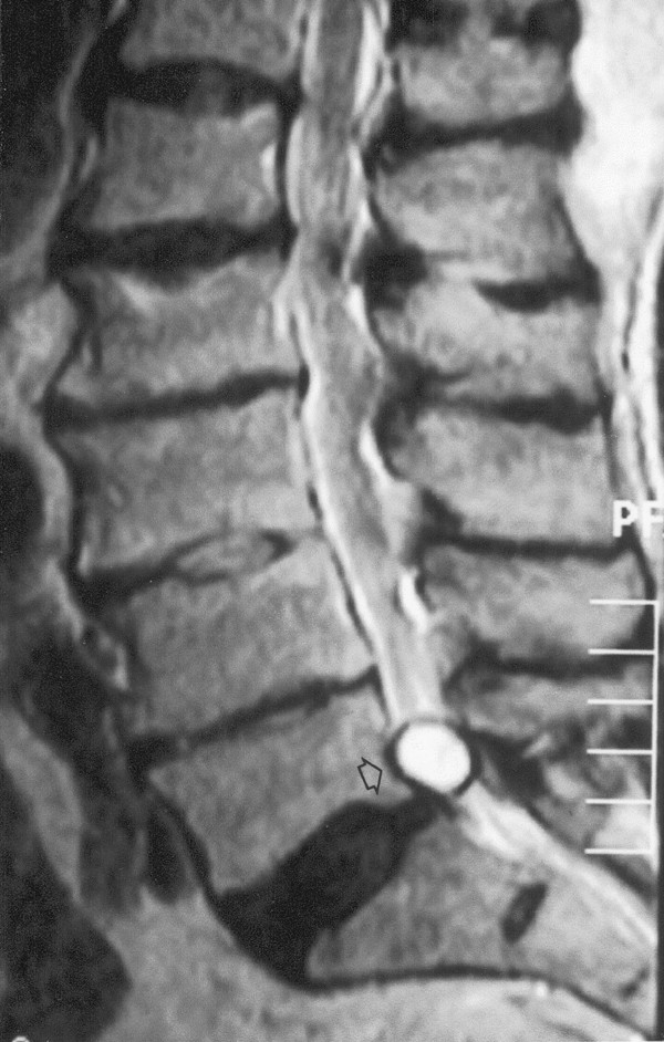
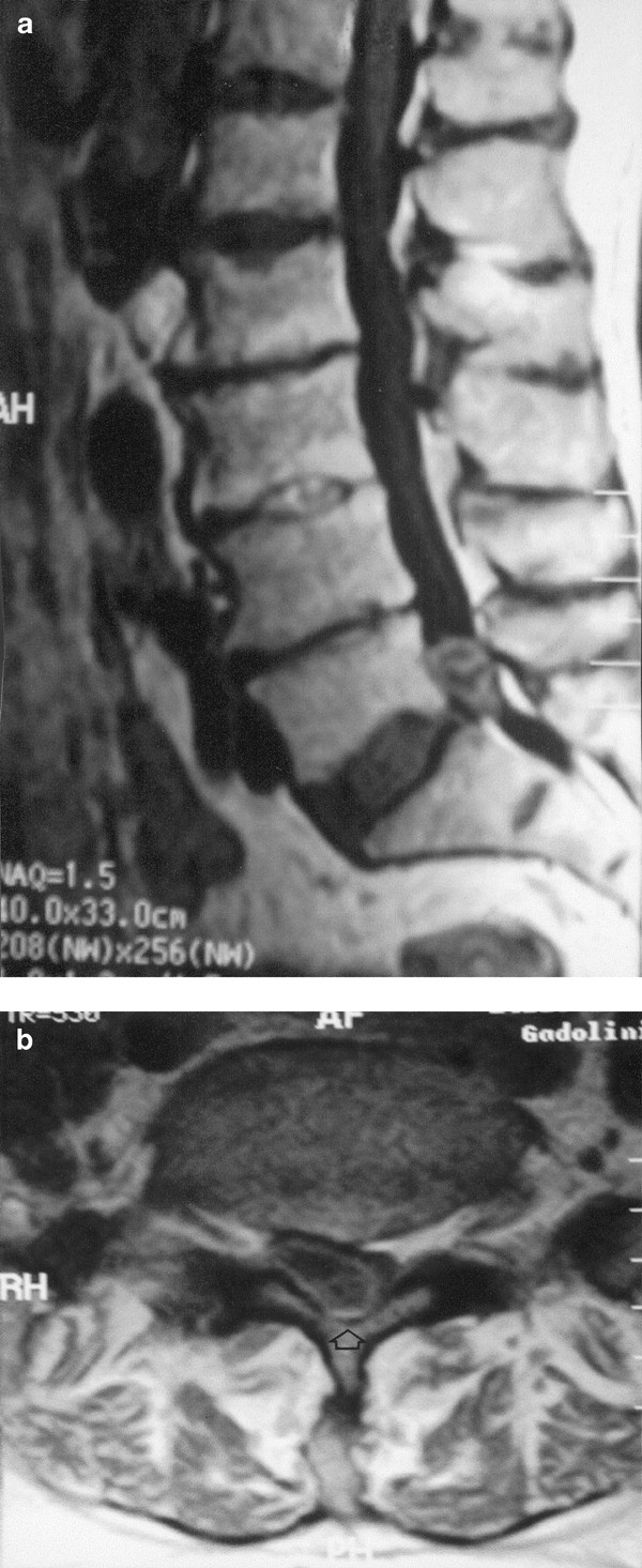
Neurological examination revealed a positive Laséque's sign, which distinguishes sciatica from disease of the hip joint, weakness of the extensor hallucis longus and decreased ankle reflex in his right lower extremity. Magnetic resonance imaging (MRI) revealed a mass-like lesion at the level of L5–S1 space. The lesion was homogenously hyperintense on noncontrast MR images, (Figure 1), which is typical for a herniated disc, but peripheral enhacement of the lesion after IV-GDPA (Figure 2) was in favor of a free disc fragment. He underwent L5 total laminectomy. The dura was swollen and immobile. A longitudinal incision was made in the dura and an intradural-free disc fragment was removed. An anterior dural rent was found communicating with the L5–S1 interspace but could not be repaired. The patient's postoperative period was uneventful and he had full recovery in 3 months.

19 × 14 mm2 hyperintense mass-like lesion on sagittal T2-weighted image (arrow)

Contrast-enhanced sagittal (a) and axial (b) images show peripherally enhancement of the lesion (arrow)
Discussion
Intradural disc herniations comprise 0.26–0.30% of all herniated discs.1 Lumbar intradural disc rupture must be considered in the differential diagnosis of mass lesions causing nerve root or cauda equina syndromes. Intradural disc herniations are usually seen at L4–L5 and L3–L4, but have also been reported at other levels. A total of 86 cases of intradural lumbar disc herniations have been reported since 1942 and only a tenth of cases occurred at L5–S1. The mechanism of intradural lumbar disc herniation and the frequent preference of L4–L5 are not well understood,3 but it is believed that adhesions between the ventral wall of the dura and posterior longitudinal ligament could act as a preconditioning factor.4 An autopsy study has shown that the ventral dura is most frequently and firmly attached to the posterior longitudinal ligament at the L4–L5 level and that these adhesions may be congenital.3
It is usually not difficult with current MRI techniques to differentiate lumbar disc herniation from other conditions.5 Contrast-enhanced MRI scans are useful to differentiate a herniated disc from a disc space infection or tumor. Peripheral enhancement around the nonenhancing disc fragment is commonly seen on contrast MRI. A herniated disc fragment will rarely enhance centrally, attributed to vascular granulation tissue infiltrating the fragment.6
In our case, the lesion was homogeneously hyperintense on T2-weighted MRI scans and that led use to suspect an intradural extramedullary tumor lesion, but on contrast MRI scans there was peripheral enhancement of the lesion, which is typical for a disc fragment.7
This case demonstrates the role and the importance of contrast MRI in the diagnosis of intradural disc herniation.
References
-
Negovetic L, Cerina V, Sajko T, Glavic Z . Intradural disc herniation at the T1–T2 level. Croat Med J 2001; 42: 193–195.
-
Dandy WE . Serious complications of ruptured intervertebral disks. JAMA 1942; 119: 474–477.
-
Yildizhan A et al. Intradural disc herniations pathogenesis, clinical picture, diagnosis and treatment. Acta Neurochir (Wien) 1991; 110: 160–165.
-
Koc RK, Akdemir H, Oktem IS, Menku A . Intradural lumbar disc herniation: report of two cases. Neurosurg Rev 2001; 24: 44–47.
-
Ramsey RG . Neuroradiology. 3rd edn. Saunders: Philadelphia 1994, 793 pp.
-
Scott WA . Magnetic Resonance Imaging of the Brain and Spine, Vol 2: LWW 2002. 3rd edn. pp 1527–1529.
-
Lidov M et al. MRI of lumbar intradural disc herniation. Clin Imaging 1994; 18: 173–178.
Author information
Authors and Affiliations
Rights and permissions
About this article
Cite this article
Aydin, M., Ozel, S., Sen, O. et al. Intradural disc mimicking: a spinal tumor lesion. Spinal Cord 42, 52–54 (2004). https://doi.org/10.1038/sj.sc.3101476
-
Published:
-
Issue Date:
-
DOI : https://doi.org/10.1038/sj.sc.3101476
Keywords
- disc
- intradural
- magnetic resonance imaging
Further reading
Source: https://www.nature.com/articles/3101476
0 Response to "Continued Herniated Mass in Nerve Root"
Post a Comment